Introduction: Review of Anatomy & Physiology of the Eye
Optician Techniques – 2nd Stage
1. Learning Objectives
By the end of this session, students should be able to:
-
>Name the main external and internal structures of the eye and state their basic functions.
>Describe how light travels through the eye to form an image on the retina.
>Explain the roles of the cornea, lens, aqueous and vitreous humors, iris/pupil, and retina in vision.
>Relate basic anatomy to common refractive errors (myopia, hyperopia, astigmatism, presbyopia).
>Use correct anatomical terms when discussing cases with colleagues or patients.
2. Why This Matters for Opticians
As an optician, you will:
-
>Take measurements that depend on eye shape and size (e.g., vertex distance, pantoscopic tilt, frame fit).
>Explain to patients how their lenses work in terms of focusing light onto the retina.
>Recognize when a problem is likely optical (needing glasses/contacts) vs. medical (needing an ophthalmologist).
A solid anatomy/physiology base makes your explanations clearer and your work more accurate. [5][8][10]
3. Big Picture: The Eye as an Optical System
Think of the eye as a camera:
-
>Cornea + lens: The camera's lens system (focus light).
>Iris/pupil: The aperture (controls amount of light).
>Retina: The film/sensor (captures the image).
>Optic nerve: The cable sending data to the brain. [5][8]
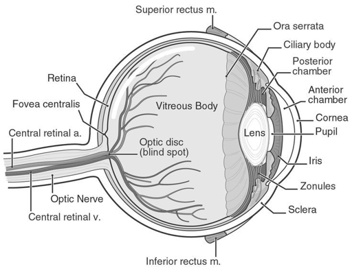
4. External Anatomy (What You See)
4.1 Orbit and Adnexa
-
>Orbit: Bony socket that protects the eyeball.
>Eyelids and lashes: Protect from dust, spread tears, limit light.
>Conjunctiva: Thin, transparent membrane covering the sclera and inner eyelids; keeps surface moist. [5][8][13]
4.2 Visible Parts of the Eyeball
-
>Sclera: White, tough outer coat; maintains shape and protects inner structures.
>Cornea: Clear, dome‑shaped front window; major refracting surface.
>Iris: Colored part; contains muscles that change pupil size.
>Pupil: Black central opening; lets light in. [5][8][10]
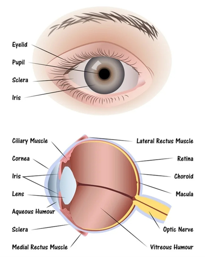
5. Internal Anatomy: Layers and Chambers
5.1 Three Tunics (Layers) of the Eyeball
-
>Fibrous tunic (outer):
-
>Cornea (front, transparent)
>Sclera (rest of outer wall, white)
Function: Protection, shape, and most of the eye's refractive power (cornea). [6][10]
-
>Choroid: Blood‑rich layer supplying retina.
>Ciliary body: Produces aqueous humor; contains ciliary muscle for accommodation.
>Iris: Controls pupil size. [6][10]
-
>Retina: Light‑sensitive layer with photoreceptors (rods and cones).
>Includes macula and fovea for sharp central vision. [6][8][10]
5.2 Segments and Chambers
-
>Anterior segment: Everything in front of the lens.
-
>Anterior chamber: Between cornea and iris – filled with aqueous humor.
>Posterior chamber: Between iris and lens – also aqueous humor.
-
>Vitreous chamber: Filled with vitreous humor (gel‑like), supports retina and maintains shape. [6][8]
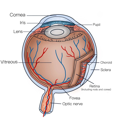
6. Key Structures and Their Functions
6.1 Cornea
Transparent, avascular (no blood vessels). Provides ~2/3 of the eye's total refractive power. Must stay clear and regularly curved for good vision; irregularities cause astigmatism. [5][8][13]
6.2 Aqueous Humor
Clear fluid produced by ciliary body. Flows: posterior chamber → pupil → anterior chamber → drains via trabecular meshwork. Functions: nourishes cornea and lens, maintains intraocular pressure (IOP). Problems with drainage → glaucoma risk. [6][8]
6.3 Iris and Pupil
-
>Iris muscles: Sphincter pupillae (constricts) and Dilator pupillae (dilates).
>Pupil size effects: Controls light entry, depth of field, and optical aberrations. [6][8]
6.4 Lens
Transparent, biconvex structure behind the iris. Suspended by zonules. Fine-tunes focus (accommodation). With age, lens stiffens → presbyopia. [6][8][14]
6.5 Vitreous Humor
Gel‑like substance filling the posterior segment. Helps maintain eye shape and keeps retina in place. [6][8]
6.6 Retina, Macula, and Fovea
-
>Retina: Contains rods (low-light) and cones (color/detail).
>Macula: Central area for detailed vision.
>Fovea: Tiny pit with highest cone density → sharpest vision. [6][8][10]
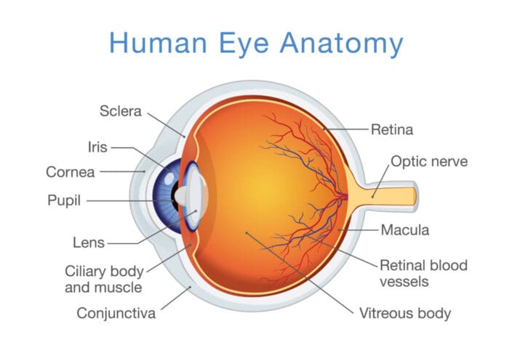
6.7 Optic Nerve
Bundle of nerve fibers carrying visual signals from retina to brain. Exits at the optic disc (blind spot). [6][8]
7. Physiology of Vision: From Light to Perception
7.1 Optical Pathway
-
>Light enters through cornea (major refraction).
>Passes through aqueous humor → pupil.
>Passes through lens (fine focusing).
>Travels through vitreous humor to the retina.
>Photoreceptors convert light into electrical signals.
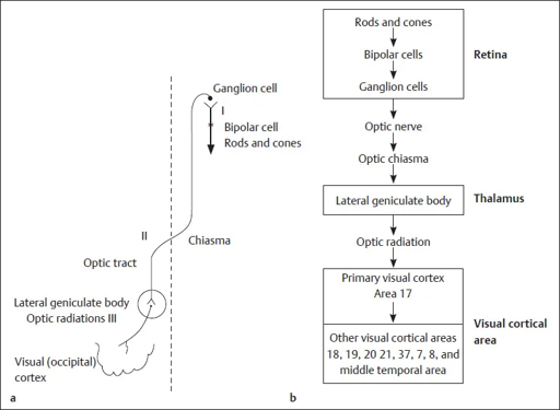
>Signals travel via optic nerve → brain → visual cortex. [5][8][27]
7.2 Accommodation
-
>Distance: Ciliary muscle relaxed → lens flatter.
>Near: Ciliary muscle contracts → lens more convex.
>Declines with age → presbyopia. [6][8]
8. Linking Anatomy to Common Refractive Errors
| Condition | What's happening optically | Typical anatomical/optical cause | Lens solution (basic idea) |
|---|---|---|---|
| Myopia (nearsighted) | Image focuses in front of retina | Eye too long or cornea/lens too powerful | Minus (−) lenses diverge light |
| Hyperopia (farsighted) | Image would focus behind retina | Eye too short or cornea/lens too weak | Plus (+) lenses converge light |
| Astigmatism | Different meridians focus at different points | Cornea or lens not perfectly spherical | Cylindrical/toric lenses |
| Presbyopia | Loss of accommodation for near | Lens stiffens, ciliary muscle less effective | Add power (reading segment, progressives) |
Sources: [5][8][14]
9. Clinical Relevance for Opticians
You don't diagnose disease, but you should recognize red flags:
-
>Cornea: Scars, keratoconus → irregular astigmatism.
>Lens: Cataract → cloudy lens, reduced acuity, glare.
>Aqueous humor / IOP: High pressure risk for glaucoma.
>Retina / macula: Sudden distortion, central blur, flashes/floaters.
10. Visual Review
11. In‑Class Activity (10–15 min)
Task: In pairs, label a blank eye diagram with: Cornea, sclera, iris, pupil, lens, aqueous humor, vitreous humor, retina, macula/fovea, optic nerve, choroid, ciliary body.
Then, write one sentence about function and one optical/clinical note for each.
12. Key Terms to Master
Cornea, sclera, conjunctiva, iris, pupil, lens, zonules, ciliary body, aqueous humor, vitreous humor, retina, rods, cones, macula, fovea, optic nerve, optic disc, anterior/posterior chamber, accommodation, refraction, intraocular pressure. [5][6][8][10]
13. Suggested Homework
-
>Draw and label the eye from memory.
>Write a short paragraph explaining how the eye focuses on a book at 40 cm vs. a sign at 5 m.
>Research one condition (e.g., keratoconus, cataract) and summarize affected structure and effect on vision.


Comments